Attention Members: Please avoid clicking on any unsolicited emails with attachments. If you have any concerns, please contact us at info@veteducation.com.au for clarification.
Attention Members: Please avoid clicking on any unsolicited emails with attachments. If you have any concerns, please contact us at info@veteducation.com.au for clarification.
Philip R Judge BVSc MVS PG Cert Vet Stud MACVSc (Vet. Emergency and Critical Care; Medicine of Dogs)
Introduction
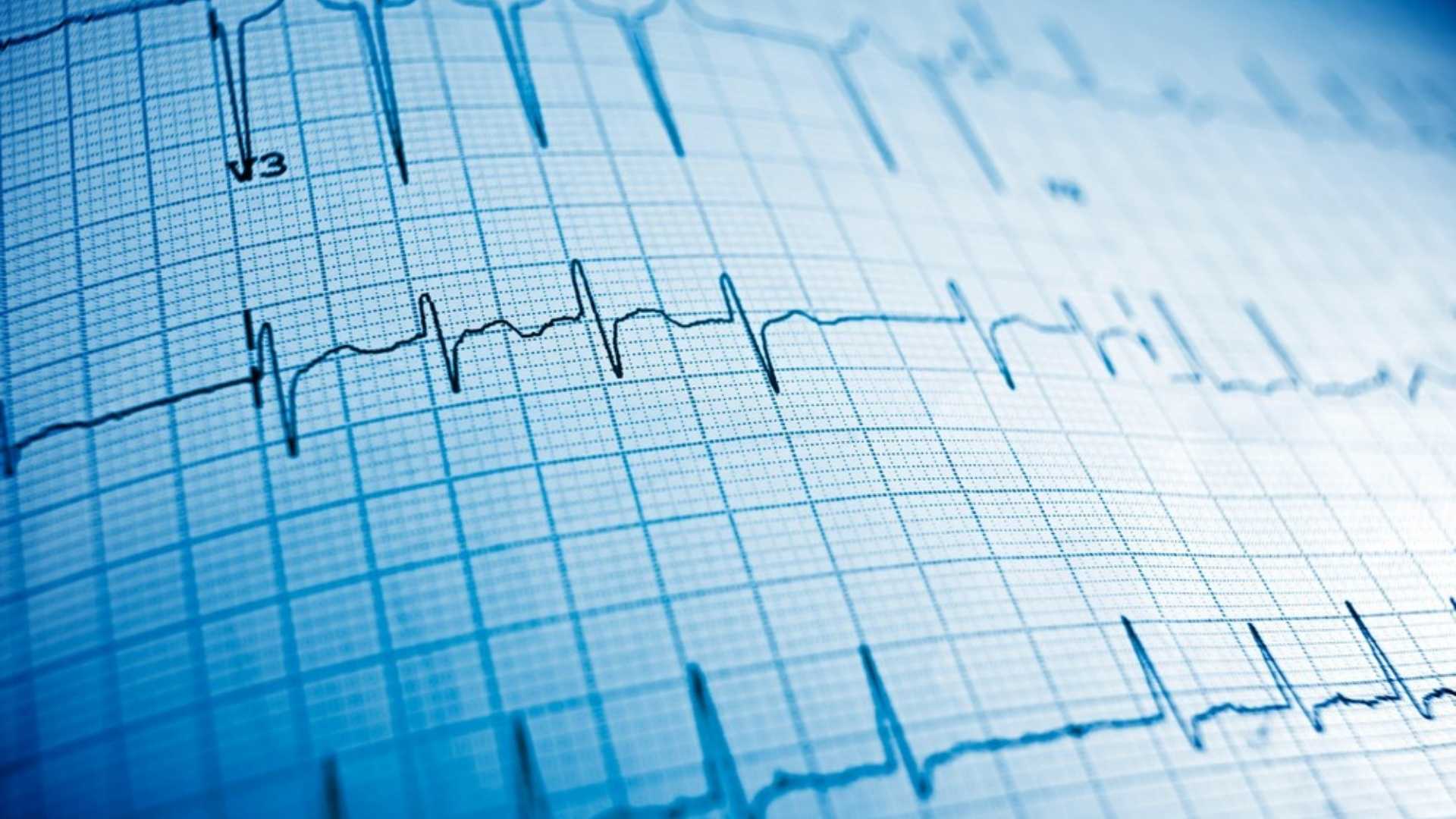
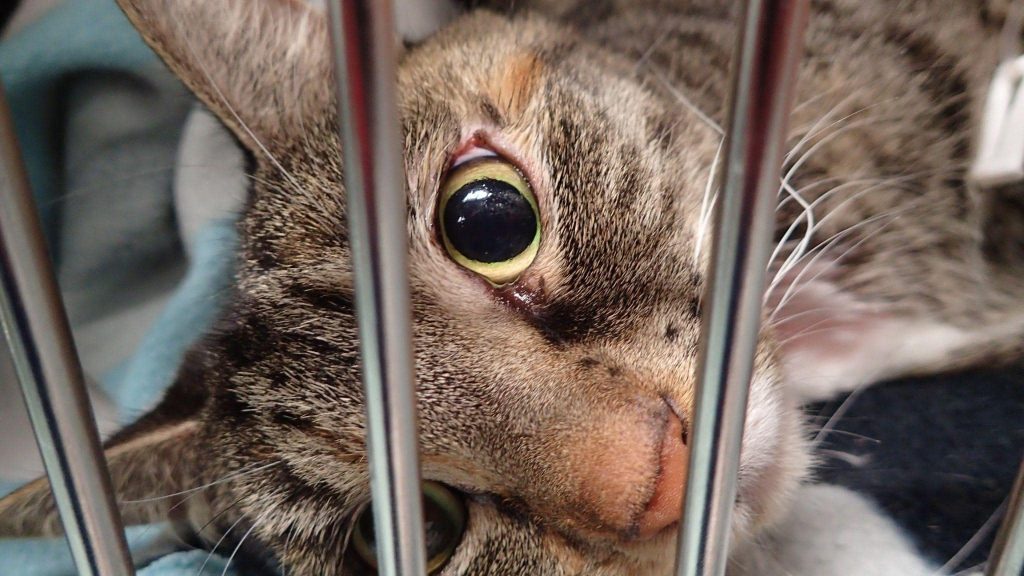
Supplementary oxygen is one of the most common interventions in both emergency and critically ill patients – used to treat hypoxia or hypoxaemia in both acute and chronic respiratory conditions, as well as part of efforts to improve oxygen delivery to tissues following trauma, injury or illness.
However, oxygen supplementation is not without risk – with oxygen toxicity being described in literature since the 1960s. The aim of this short review is to assess literature surrounding oxygen therapy and the potential for oxygen toxicity in both emergency and critically ill dogs and cats.
Mechanisms of Oxygen Toxicity
Oxygen is a highly reactive element. In the body, oxygen molecules can be converted to “reactive oxygen species” – meaning oxygen molecules or oxygen species that can gain or lose electrons, with resultant potential to damage cells and tissues in the process1,2.
The most common reactive oxygen species include1,2
Superoxide anion (O2–)
Hydrogen peroxide (H2O2)
Hydroxyl radicals (OH–)
Singlet oxygen (O–)
Hypochlorous anion (HOCl–)
Ozone (O3)
Reactive oxygen species cause structural damage to lipid membranes, proteins and nucleic acids in cells. This damage leads to altered and disordered cell metabolism and function, and ultimately cell dysfunction, injury and cell death1,2. The most biologically significant of these reactive oxidant species are the hydroxyl ion and peroxynitrite. Peroxynitrite is the product of the reaction between superoxide and nitric oxide, and interacts with lipids, DNA, and proteins via direct oxidative reactions or via indirect, radical-mediated mechanisms2. These reactions trigger cellular responses ranging from subtle modulations of cell signaling to overwhelming oxidative injury2,3.
Under normal circumstances, less than 5% of cellular oxygen is converted to reactive oxygen species, and these are rapidly removed from cell by endogenous scavengers, or antioxidants, including1,2:
Superoxide dismutase
Metalloproteins
Catalase
Glutathione peroxidase
Glutathione reductase
Vitamin C
Vitamin E
Beta-carotene
Uric acid
Excessive oxygen supplementation can disrupt the balance between reactive oxygen species production, and free radical scavengers, leading to an excess of reactive oxygen species – ultimately increasing the potential for cell and organ damage1.
The Early Evidence for Oxygen Toxicity
There are numerous published studies demonstrating the adverse effects of excess oxygen – or hyperoxia – in both humans and animals, and in both experimental and clinical settings. It was reported in the early 1970s that breathing 50-100% oxygen at one atmosphere was potentially toxic to the lungs. Subsequently, there is recognition of the toxic effects on other systems of the body including the eyes, red cells, liver, heart, kidneys and endocrine systems as well as general damage to cells2,3.
Early studies on pulmonary oxygen toxicity revealed interesting findings, including1
That an FiO2 of greater than 60% was associated with lung injury
That FiO2 between 50% and 60% was generally well-tolerated for prolonged periods
That patients maintained on high FiO2 were more likely to suffer lung injury if they were subjected to elevated oxygen concentrations for longer periods of time
There is considerable individual variation in susceptibility to lung injury following periods of hyperoxia
Animals that survived exposure to high oxygen concentrations showed some ability to adapt/acclimate
The Pathology of Lung Damage Resulting from Oxygen Toxicity
The injury caused by hyperoxia is similar to that of acute respiratory distress syndrome, and includes the development of pulmonary oedema, diffuse alveolar membrane damage, inflammation of the respiratory mucosa, pulmonary consolidation, and pleural fluid accumulation1,2.
Clinically, these changes are manifested as increased respiratory effort, bilateral crackles in thoracic auscultation, and haemorrhagic, frothy sputum and heamoptysis1,2.
Radiography typically reveals diffuse pulmonary infiltrates in an alveolar/interstitial and bronchial pattern1,2.
Pulmonary oxygen toxicity is characterised by an initial latent, or lag period in which no overt clinical manifestations of toxicity can be detected. The duration of this latent period is inversely proportional to the level of inspired oxygen, and can vary from 4 to 22 hours in patients receiving greater than 95% oxygen2. In injured or diseased lungs, in patients on 100% oxygen, symptoms may begin as early as 3 hours after exposure to oxygen.
The pattern of lung injury associated with excessive oxygen supplementation occurs as follows2,3:
Reduced trachea-bronchial cilia activity (3 hrs. on 95% inspired oxygen)
Decreased vital lung capacity
Atelectasis (24 hrs. on 100% oxygen)
Diffuse alveolar damage (24 hrs. or longer on >50% oxygen)
What Recent Evidence is there for Oxygen Toxicity?
Recent studies of hyperoxia in humans have focused on specific groups of patients during resuscitation from disease, including the following:
Neonatal resuscitation1,2
Higher FiO2 resuscitation (100% oxygen) is associated with higher mortality in the neonatal resuscitation period
Patients resuscitated with FiO2 concentrations of 21% (room air) had up to 30% better survival rates than those resuscitated with higher oxygen concentrations
Lower FiO2 during resuscitation reduced risks of chronic lung disease and markers of oxidative injury
Adult resuscitation in acute and intensive care1,3
Conservative oxygen therapy (SpO2 target 94-98% versus 97-100%) was associated with an 8.6% reduction in mortality in an ICU population
ICU patients with hyperoxia have a higher incidence of adverse events
In a study of over 6,000 acutely unwell patients found an increase in mortality rate of 25% for every 1% increase in SpO2
FiO2 of greater than 0.52 (52% inspired oxygen) was found to be associated with a higher 30-day mortality rate
Hyperoxaemia with a PaO2 of greater than 100 mm Hg is associated with higher mortality
Cardiac Arrest1
PaO2 in excess of 300 mm Hg is associated with higher mortality in post-cardiac arrest patients
Functionality is worse in patients resuscitated with hyperoxia
Traumatic Brain Injury1
Patients with severe traumatic brain injury that were resuscitated with PaO2 concentrations of greater than 200 mm Hg had worse discharge coma scores, and higher mortality than patients resuscitated with a PaO2 of between 100-200 mm Hg
Other organs2,3
Oxidative damage secondary to hyperoxia can occur in any cell in the body, and has been implicated in red blood cell destruction, myocardial cell damage, endocrine tissue damage, and acute kidney injury
Management and Prevention of Oxygen Toxicity
In veterinary medicine, no guidelines are currently available for prevention of oxygen toxicity in acutely ill, or critically ill patients, other than suggestions to avoid excessive oxygen supplementation to reduce the potential for oxygen toxicity.
In human medicine, the Australian and New Zealand Thoracic Society has recommended target oxygen saturations of 92-95% for patients not at risk of hypercapnoeic respiratory failure1.
In light of clinical evidence, it seems reasonable to attempt to achieve a target PaO2 concentration of close to 100 mm Hg in the majority of patients requiring oxygen supplementation, and to avoid excesses above this level, in order to avoid unintended consequences of hyperoxaemia1,4.
Furthermore, judicious and appropriate use of intravenous fluid therapy, cardiac inotropes and other medications, including vasopressors (where appropriate) should be considered an essential component of patient treatment, focused on optimizing tissue oxygen delivery to vital organs, with the precise composition of therapeutic agents applied made on a case-by-case basis1,3,4.
References:
Reidy BTG, Whyte P, Neligan PJ. Is Oxygen Toxic in Evidence-Based Practice of Critical Care; Deutschman/Neligan (Ed); Elsevier (2020) P36-42.
Thomson, Louise, and James Paton. “Oxygen toxicity.” Paediatric respiratory reviews 15, no. 2 (2014): 120-123.
Hedenstierna, Göran, and Christian S. Meyhoff. “Oxygen toxicity in major emergency surgery—anything new?” Intensive Care Medicine 45, no. 12 (2019): 1802-1805.
Siemieniuk, Reed AC, Derek K. Chu, Lisa Ha-Yeon Kim, Maria-Rosa Güell-Rous, Waleed Alhazzani, Paola M. Soccal, Paul J. Karanicolas et al. “Oxygen therapy for acutely ill medical patients: a clinical practice guideline.” Bmj 363 (2018).
Learn all about the the basic principles of genetic testing, sample collection, reliable database information, test result interpretation, and recommendations for clients.
This article will explore the presentation, pertinent pathophysiology, patient selection, treatment considerations and outcomes of medical management of canine pyometra.
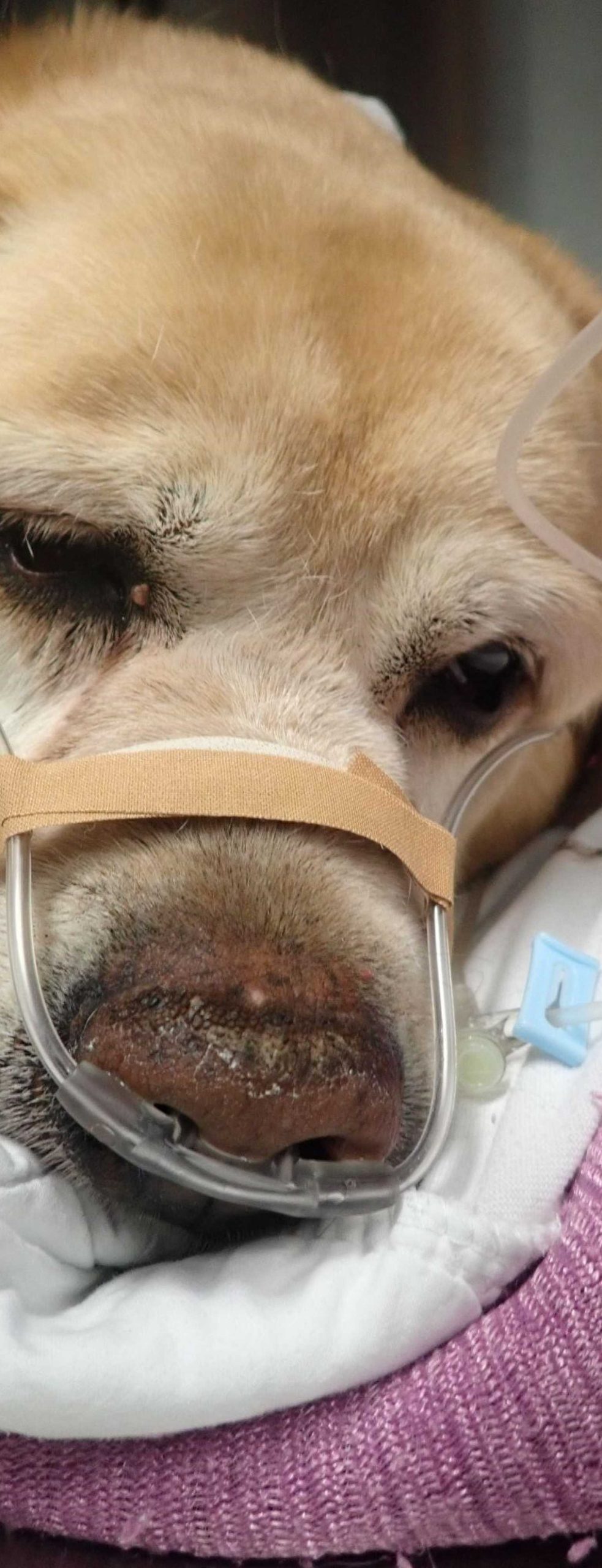
This short review will focus on the concept of oxygen toxicity, and its relevance in the treatment of the trauma patient - many of whom require and are given oxygen therapy as part of their treatment regime.