Attention Members: Please avoid clicking on any unsolicited emails with attachments. If you have any concerns, please contact us at info@veteducation.com.au for clarification.
Attention Members: Please avoid clicking on any unsolicited emails with attachments. If you have any concerns, please contact us at info@veteducation.com.au for clarification.
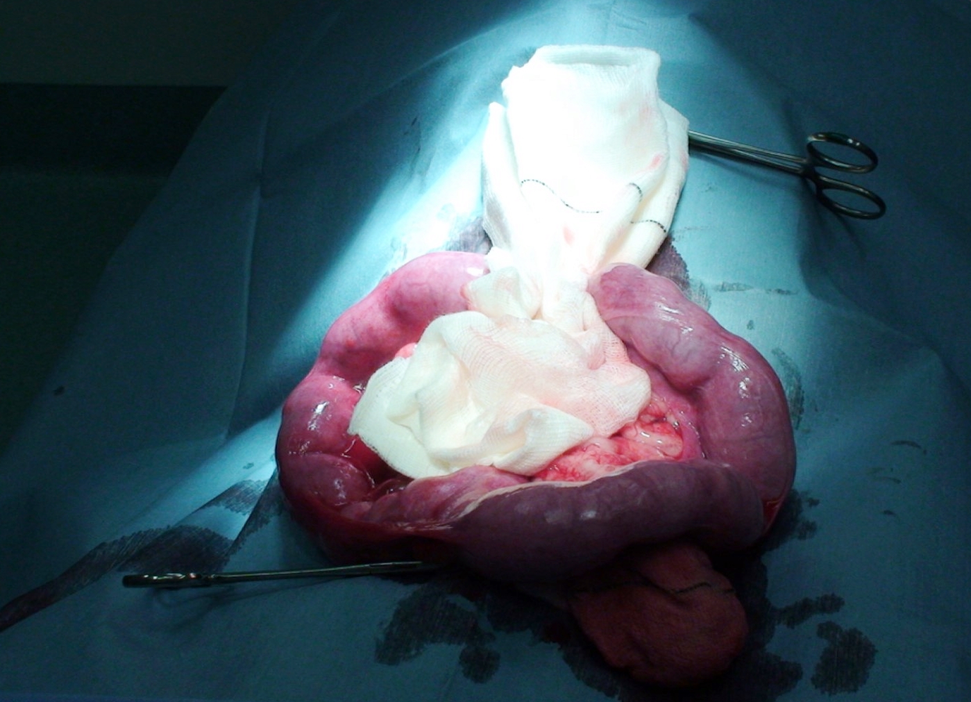
Canine pyometra is a life-threatening disease characterised by suppurative inflammation and accumulation of exudates within the uterus.1 Historically, ovariohysterectomy was the only treatment option, and today it remains the treatment of choice. However, in recent years, medical approaches have shown success and may be considered for patients that possess high genetic value or those that are high-risk anaesthetic candidates. Careful consideration must be given regarding the medical treatment approach. It requires a thorough understanding of the pathophysiology of pyometra and demands judicious patient selection. Clients must be well counselled regarding the prognosis for breeding (where applicable) and the likelihood of recurrence. This article will explore the presentation, pertinent pathophysiology, patient selection, treatment considerations and outcomes of medical management of canine pyometra.
What are the risk factors for pyometra in dogs?
Pyometra occurs most frequently in sexually mature female dogs greater than 4 years of age.2 Incidence increases with age and repeated oestrus cycles, with an average age at diagnosis of 7 years.3,4 Nulliparous intact female dogs are 6 times more likely to develop pyometra when compared with primiparous or multiparous dogs.5 Many studies suggest a genetic component, with certain breeds more commonly affected, but 25% of intact female dogs develop pyometra by the age of 10; therefore, one should consider all breeds at significant risk.4,6
What is the pathophysiology of pyometra?
Pyometra is considered a diestral disease which comprises both hormonal and bacterial components. Progesterone stimulates the proliferation of uterine glands, closes the cervix, suppresses myometrial activity and suppresses local uterine immune responses.4 Proliferation of endometrial glands leads to cystic endometrial hyperplasia, which is believed to predispose to pyometra but is not a prerequisite.
Pyometra results from the translocation of vaginal bacteria through the cervix into a progesterone-primed uterus, and it often leads to systemic inflammation and sepsis.7,8Escherichia coli is the most common bacterial isolate and accounts for more than 57–90% of cases.9,10 Other less common isolates include Staphylococcus spp., Pseudomonas spp., Proteus spp. and Klebsiella spp.10 While the pathogenesis has recently come under debate, researchers generally agree that hormonal imbalances affect epithelial cells in the uterus, which aids adherence and colonisation of bacteria.1,8,10 Culture results from up to 25% of cases are either a mixed bacterial population or culture negative.10 Negative culture results do not rule out a bacterial infection.
Diagnosis
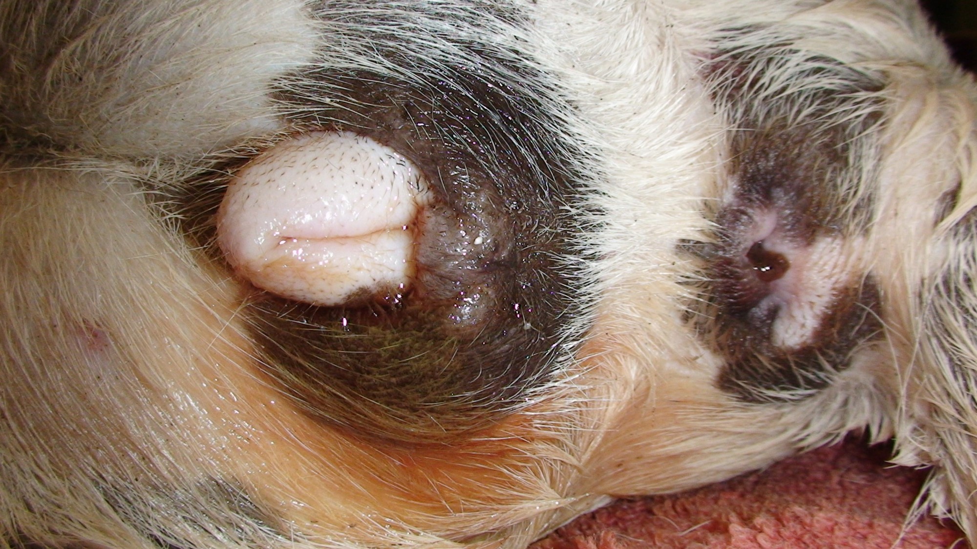
The most typical presentation is that of a middle-aged or older intact female dog 2–4 months following oestrus.10 Often, a purulent mucohaemorrhagic vaginal discharge is present, but this may be absent in the case of a closed cervix. Generally, but not always, open-cervix pyometra cases present with milder clinical signs.7 Eighty percent of all pyometra cases present dehydrated and hypoperfused.11 Other systemic effects include pyrexia, hypo- or anorexia, lethargy, polyuria/polydipsia, tachycardia and weak pulses.10 Vomiting and diarrhoea (15–30% of cases) are occasionally the owner’s primary reasons for bringing their dog to the veterinary clinic. These cases can be easily misdiagnosed without a thorough history, genital examination and laboratory analysis.
Laboratory findings generally include leukocytosis, neutrophilia – with or without a left shift, monocytosis, thrombocytopenia and azotaemia.10 Concomitant cystitis is common, however diagnosis with the aid of cystocentesis is not recommended in cases of suspected pyometra due to the risk of puncturing the distended and often friable uterus.7
Pyometra-induced uterine enlargement must be differentiated from mucometra and hydrometra.1 Both of these conditions occur due to a build-up of either sterile mucous or serous fluid, respectively.1 Generally, dogs that are affected by mucometra or hydrometra appear systemically well, but these conditions do affect fertility and, therefore, require intervention also.
Ultrasonographic imaging of the urogenital tract is an excellent tool for diagnosis. Uteromegly, thickening of the uterine walls and fluid-distension of the tubular horns are evident ultrasonographically.7 Uterine fluid is usually homogenous or flocculent, helping to differentiate it from other causes of uteromegaly. Other causes of uteromegly not already mentioned include early pregnancy, hematometra and uterine torsion.7
What are the contraindications for medical management of pyometra?
Medical management is contraindicated in the following scenarios:
Pyrexic patients suspected to have peritonitis
Systemically unwell dogs with closed pyometra – these patients should proceed to surgery shortly after initial stabilisation
Dogs with ultrasound evidence of uterine wall thinning and copious uterine content – these patients are at risk of uterine rupture or leakage and subsequent peritonitis
What are the indications for medical management of pyometra?
Medical management is preserved for the following scenarios:
Relatively young (<6 years), otherwise healthy, dogs that are genetically valuable breeding animals
Dogs that are otherwise stable but may have a medical condition that precludes immediate surgery
Dogs that require stabilisation of concurrent disease before becoming anaesthetic candidates for later ovariohysterectomy
Treatment approach
Regardless of the treatment approach, dogs presenting with systemic signs of illness or shock must be appropriately stabilised before direct treatment of uterine disease. Goals include restoration of fluid volume and perfusion pressure, correction of electrolytes, and initiation of antimicrobial therapy. Various biomarkers can be used to monitor and predict patient outcomes in critically ill patients. The patient should be hospitalised for the duration of therapy to allow close monitoring of the patient’s status and for observation of adverse effects associated with drug therapy.
Medical management protocols vary, but all share the same goals:
Stabilise the patient
Eliminate the effects of progesterone on the reproductive tract by either inducing luteolysis or antagonising progesterone binding12
Induce uterine contractions
Manage bacterial infection via appropriate antimicrobial therapy
Eliminating the effects of progesterone is achieved by using progesterone receptor antagonists (aglepristone), dopamine agonists and/or prostaglandins. Eliminating the effects of progesterone aids cervical relaxation, permits uterine contractions, improves uterine local immunity and allows uterine expulsion of purulent contents.
What drugs eliminate progesterone effects and induce uterine contractions?
Aglepristone
Aglepristone is a progesterone receptor antagonist, which is often used in combination with prostaglandins due to synergistic effects. Unlike prostaglandins, aglepristone does not have ecbolic activity and is therefore safe to use in cases of closed-cervix pyometra.7 Aglepristone at a dose of 10 mg/kg SC on days 1,2 and 8 (+/- day 15 if required) will induce cervical opening within 48 hours.13 The addition of cloprostenol, following cervical opening, at a dose of 2 µg/kg SC on days 3–7 increases the overall success rate from 60% to greater than 80%.13
Prostaglandins
Multiple prostaglandin protocols exist, but lower doses at higher frequencies appear to mitigate adverse effects with the most success. Dinoprost is a natural prostaglandin and is given at 100–250 µg/kg/day divided into 3 to 6 doses over 24 hours for up to 10 days, or until the resolution of vaginal discharge.7 It is recommended to start at the lower end of the dosing range and increase to the higher end over subsequent days while observing for adverse effects – panting, emesis, hypersalivation, anxiety, shivering, diarrhoea and urination.7 Extreme care must be taken with brachycephalic breeds as they may be predisposed to bronchospasm.10 Cloprostenol, a synthetic prostaglandin, arguably is accompanied by fewer side effects and is given at a dose of 1–5 µg/kg SC every 24 hours. 7
Dopamine Agonists
Dopamine agonists, such as cabergoline or bromocriptine, have significant anti-prolactin activity (and therefore anti-luteotropic activity). When used in combination with prostaglandins, serum progesterone levels decline in as little as 24 hours.12 Cabergoline is often preferred because it requires only once-daily dosing, as opposed to bromocriptine, which requires dosing three times daily. Cabergoline is dosed at 5µg/kg PO q24 hours, or the dose can be divided and given once every 12 hours to avoid gastrointestinal upset.7 The drug is given alongside prostaglandins until serum progesterone declines to less than 3.2 nmol/L (1ng/ml) for up to 14 days. Success rates of combined cabergoline/cloprostenol protocols vary within the range of 80%–90%.14,15 Cabergoline is available in 2mg, 1mg and 0.5mg tablets and, therefore, is best suited for large dogs, or must otherwise be compounded. For this reason, in countries where aglepristone is available, protocols that utilise aglepristone may be preferred.
Antimicrobial therapy
Samples for culture and sensitivity should be collected from the anterior vagina using a double-guarded swab or, if available, transcervical endoscopy. Because E. coli is the primary bacteria isolated from up to 90% of canine pyometra cases, initial empirical antimicrobial therapy should cover E.coli. The clinician must consider that endotoxemia may occur following E. coli death and subsequent release of lipopolysaccharide into systemic circulation.
While culture and sensitivity results are pending, amoxicillin-clavulanate or a combination of penicillin and fluoroquinolone are good first options. For dogs that initially experience emesis due to prostaglandin use, injectable antimicrobial therapy may be considered. Many clinicians use ampicillin 10–20 mg/kg IV q6h +/- enrofloxacin. Amoxicillin-sulbactam 10–20 mg/kg IV q8h administered slowly over 15–20 mins has been used empirically with success, but it is argued that this drug should be reserved for cases of sepsis.
Patient monitoring
Serum progesterone levels should be monitored daily to confirm they are declining and the patient is responding to therapy. Serum progesterone greater than 6.3 nmol/L (2ng/ml) indicates that functional corpus luteum (CL) tissue is still present.
Intravenous fluid therapy should continue until hydration and perfusion are corrected, serum electrolyte values return to normal, acid–base status normalises, and emesis is controlled. Antiemetics may be given to prevent prostaglandin-induced emesis.
Transabdominal ultrasound is performed daily to monitor uterine size and evacuation of luminal contents. Clinical improvement is expected within 48 hours of starting therapy. Resolution of pyometra involves complete uterine evacuation, cessation of vaginal discharge, attainment of baseline serum progesterone concentration (<3.2 nmol/L; <1 ng/ml), normalisation of haematological and biochemical parameters and a return to normal diet and activity. Antimicrobial therapy should continue for two weeks beyond the complete resolution of pyometra.12
What is the prognosis for pyometra following surgical versus medical management?
The prognosis for survival in appropriately selected cases is good, while the prognosis for fertility varies (14–92%).7,10,13,16 Fertility in younger dogs is generally better than that of older dogs.7 Pyometra recurs in approximately 20% of cases, and this must be stressed to the owner before beginning medical treatment.17 Pregnancy is generally considered protective; therefore, the female dog should either be bred on her next oestrus cycle (with cranial vaginal culture and sensitivity being performed at proestrus) or undergo ovariohysterectomy beforehand.18
Key Points
Canine pyometra is a life-threatening disease characterised by suppurative inflammation and accumulation of exudates within the uterus.1
Medical approaches have shown success and may be considered for patients that possess high genetic value or those that are high-risk anaesthetic candidates.
Medical protocols aim to stabilise the patient, eliminate the effects of progesterone on the reproductive tract, induce uterine contractions and manage bacterial infection.
Eliminating the effects of progesterone is achieved by using progesterone receptor antagonists (aglepristone), dopamine agonists and/or prostaglandins.
The prognosis for survival in appropriately selected cases is good, while the prognosis for fertility varies (14–92%).7,10,13,16
Pregnancy is generally considered protective; therefore, the female dog should either be bred on her next oestrus cycle or undergo ovariohysterectomy beforehand.18
References
Santana CH, Santos RL. Canine pyometra – an update and revision of diagnostic terminology. Brazilian Journal of Veterinary Pathology 2021;14:1–8.
Smith FO. Canine pyometra. Theriogenology 2006;66:610–612.
Jitpean S, Ström-Holst B, Emanuelson U, et al. Outcome of pyometra in female dogs and predictors of peritonitis and prolonged postoperative hospitalization in surgically treated cases. BMC veterinary research 2014;10:1–12.
Jitpean S, Hagman R, Ström Holst B, et al. Breed variations in the incidence of pyometra and mammary tumours in Swedish dogs. Reprod Domest Anim 2012;47 Suppl 6:347–350.
Niskanen M, Thrusfield M. Associations between age, parity, hormonal therapy and breed and pyometra in Finnish dogs. Veterinary Record 1998;143:493-498.
Egenvall A, Hagman R, Bonnett BN, et al. Breed risk of pyometra in insured dogs in Sweden. J Vet Intern Med 2001;15:530–538.
Wallace GBaC, M.L. A review of pyometra in small animal medicine: incidence, pathophysiology, clinical diagnosis, and medical management Clinical Theriogenology 2018;10:435–452.
Fieni F, Topie E, Gogny A. Medical treatment for pyometra in dogs. Reproduction in domestic animals 2014;49:28–32.
Lopes CE, De Carli S, Riboldi CI, et al. Pet Pyometra: Correlating bacteria pathogenicity to endometrial histological changes. Pathogens 2021;10:833.
Hagman R. Pyometra in small animals 2.0. Vet Clin North Am Small Anim Pract 2022;52:631–657.
Børresen B. Pyometra in the dog. II – A pathophysiological investigation. II. Anamnestic, clinical and reproductive aspects. Nord Vet Med 1979;31:251–257.
Verstegen J, Dhaliwal G, Verstegen-Onclin K. Mucometra, cystic endometrial hyperplasia and pyometra in the bitch: advances in treatment and assessment of future reproductive success. Theriogenology 2008;70:364–374.
Fieni F. Clinical evaluation of the use of aglepristone, with or without cloprostenol, to treat cystic endometrial hyperplasia-pyometra complex in bitches. Theriogenology 2006;66:1550–1556.
England GC, Freeman SL, Russo M. Treatment of spontaneous pyometra in 22 bitches with a combination of cabergoline and cloprostenol. Vet Rec 2007;160:293–296.
Corrada Y, Arias D, Rodríguez R, et al. Combination dopamine agonist and prostaglandin agonist treatment of cystic endometrial hyperplasia – pyometra complex in the bitch. Theriogenology 2006;66:1557–1559.
Melandri M, Veronesi MC, Pisu MC, et al. Fertility outcome after medically treated pyometra in dogs. Journal of veterinary science 2019;20.
Gobello C, Castex G, Klima L, et al. A study of two protocols combining aglepristone and cloprostenol to treat open cervix pyometra in the bitch. Theriogenology 2003;60:901–908.
Hagman R, Lagerstedt A-S, Hedhammar Å, et al. A breed-matched case-control study of potential risk-factors for canine pyometra. Theriogenology 2011;75:1251–1257.
Learn all about the the basic principles of genetic testing, sample collection, reliable database information, test result interpretation, and recommendations for clients.
This article will explore the presentation, pertinent pathophysiology, patient selection, treatment considerations and outcomes of medical management of canine pyometra.
This short review will focus on the concept of oxygen toxicity, and its relevance in the treatment of the trauma patient - many of whom require and are given oxygen therapy as part of their treatment regime.