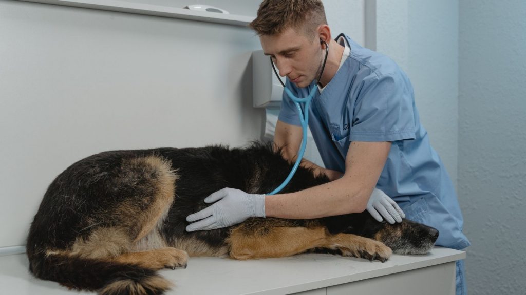
This short write-up aims to review some of the recent research being conducted on the use of biomarkers – both for the identification of sepsis and in attempting to predict survivors from non-survivors in critical illness.
The search for the ability to ‘predict’ the likely outcome in a critically ill patient long before their actual outcome has been going on for several years. Having this ability has important ramifications for not only the patient, who must endure their illness and treatment interventions, but also for the client – who must pay sometimes significant amounts of money to pay for treatment of their critically ill pet; and also for veterinary hospitals, who invest significant resources in the care of these patients.
Several studies have been conducted in dogs and cats to determine the diagnostic and prognostic utility of serum biomarkers in patients with severe illness. As yet, there are no confirmed, reliable biomarkers able to detect survivors from non-survivors at the time of diagnosis. However, trends in several biomarkers may help distinguish survivors from non-survivors over time.
Sepsis and systemic inflammatory response (SIRS) are associated with increased production of the proinflammatory cytokines TNF and IL-6. In addition, sepsis is associated with decreased production of the anti-inflammatory cytokine IL-10.
So, let’s look at some of the literature surrounding these biomarkers, and see what has been found.
The first article we will review is an original study by Gommeren and colleagues,[1] published in JVECCS in January of 2018 – looking at inflammatory cytokine and C-reactive protein concentrations in dogs with systemic inflammatory response syndrome.
Sixty-nine dogs with a diagnosis of systemic inflammatory response syndrome were included in the study. The dogs were diagnosed with SIRS using the classic SIRS criteria, which include elevated or depressed body temperature, elevated or depressed white blood cell count, elevated heart rate and slow respiratory rate – with a dog meeting any 2 of these criteria being eligible for inclusion in subsequent analysis.
Blood was taken at presentation, then at 6hr, 12 hrs., 24 hrs. and 72 hrs. following presentation, and at least 1 month following discharge.
Dogs had diseases including gastric dilatation-volvulus (GDV), infection, neoplasia, trauma, kidney disease, gastroenteritis and a range of other conditions.
Blood collected was tested for the following:
- C-reactive protein
- Interleukin-6
- Tumour necrosis factor-alpha
Serum C-reactive protein concentration was found to be increased in dogs with clinical signs of SIRS, and was positively correlated with increased plasma concentrations of interleukin-6
However, neither absolute values, nor changes in blood CRP, IL-6 or THF-alpha concentrations helped to identify the underlying disease, or to predict outcome in this cohort of dogs.
The second article we will look at is by Casey Kohen and colleagues,[2] published in the same issue of JVECCS as the last article of January 2018.
This article presents a retrospective evaluation of the prognostic utility of plasma lactate concentration, base deficit, pH, and anion gap in canine and feline emergency patients.
The authors reviewed the records of 566 dogs and 185 cats presenting to the emergency room at UC Davis, that had venous blood gases and blood lactate performed within 2 hours of presentation.
Patients were admitted for a number of reasons, including
- Trauma
- Haemorrhage
- Sepsis
- Neoplasia
- Respiratory disease
- Cardiac disease
- Hepatic disease
- GI disease
- Urinary tract disease
- Endocrine or reproductive disease
- Neurological disease
- Haematological disease
- Toxicosis
Cats with lactic acidosis had the highest mortality (49%); versus cat with hyperlactataemia and no acidosis (44.6%), or normal lactate (22.6%).
Dogs with lactic acidosis had a mortality rate of 59.8%; whereas the mortality rate in dogs with hyperlactataemia and normal blood gas analysis, along with those with normal lactate were not significantly different, at around 24%.
Results of this study indicated that:
- Median plasma lactate concentration and anion gap were higher, and base deficit was more negative, and pH was lower in non-survivors than in survivors.
- Blood pH, plasma lactate, and base deficit were independent predictors of mortality in dogs
- Only plasma lactate was an independent predictor of mortality in cats
The study concludes that in addition to the above-mentioned findings that the presence and magnitude of hyperlactataemia on presentation to the ER may help identify dogs and cats with a high likelihood of in-hospital mortality, particularly if present with acidosis. This means that evaluation of blood gas, along with blood lactate may have more value than testing blood lactate alone.
This study did not look at subsequent lactate readings over time, to determine if the rate of decline in lactate offered greater sensitivity, and this is an important area of further research that is recommended.
In another study of 45 dogs – 22 with sepsis and 23 with non-infectious systemic inflammation[3], concentrations of TNF, IL-6, IL-10 and CXCL-8 (CX Chemokine Ligand-10) were measured and analysed against survival, and ability to differentiate non-infectious SIRS from sepsis. None of the biomarkers had optimal sensitivity or specificity for diagnosis of sepsis and none were predictive of outcome.
Another study conducted using 61 dogs with sepsis and non-infectious SIRS[4], C-reactive protein (CRP) was measured. There was no significant relationship between the survival rate in dogs with non-septic SIRS or sepsis and the initial serum CRP concentrations. However, over the 3-day study period, surviving dogs displayed a significantly greater decrease in CRP than non-survivors, and this reduction correctly predicted survival in 94% of dogs and death in 30% of the dogs, with a false positive rate of 22%.
In another study of 20 dogs with sepsis[5], compared to 28 control non-septic dogs, protein C (PC), antithrombin (AT), pro-thrombin (PT), partial thromboplastin times (PTT), fibrinogen degradation products (FDP) and D-dimers (DD) were measured within 24-hours of admission to hospital.
PC and AT activities were significantly lower in dogs with sepsis compared to controls. Additionally, dogs with sepsis had significantly higher PT, PTT, D-dimer, and FDP compared to controls. Platelet counts were not significantly different between groups. Ten of the 20 septic dogs (50%) died, but no association was identified between any of the measured variables and outcome.
A subsequent study[6] showed that PC and AT activities changed significantly over time in dogs with sepsis and increasing concentrations of both over the course of hospitalization for sepsis is likely related to survival.
What do these studies tell us?
All of these studies contribute to, and build on our understanding of the predictive values of cytokines and metabolites in patients with severe illness. Based on this short review of the literature, blood lactate, in conjunction with blood gas analysis, as well as trends in concentrations of protein C and antithrombin, may provide some insight into survivors versus non-survivors. Further research is underway in this interesting field that will hopefully help us, as clinicians in the field, best direct our treatment and resuscitation efforts, whilst assisting decision-making processes for our clients and their beloved pets.
References:
- Gommeren K, Desmas I, Garcia A, Bauer N, Moritz A, Roth J, Peeters D. Inflammatory cytokine and C‐reactive protein concentrations in dogs with systemic inflammatory response syndrome. Journal of veterinary emergency and critical care. 2018 Jan;28(1):9-19.
- Kohen CJ, Hopper K, Kass PH, Epstein SE. Retrospective evaluation of the prognostic utility of plasma lactate concentration, base deficit, pH, and anion gap in canine and feline emergency patients. Journal of veterinary emergency and critical care. 2018 Jan;28(1):54-61.
- DeClue AE, Sharp CR, Harmon M. Plasma inflammatory mediator concentrations at ICU admission in dogs with naturally developing sepsis. Journal of veterinary internal medicine. 2012 May 1;26(3):624-30.
- Gebhardt C, Hirschberger J, Rau S, Arndt G, Krainer K, Schweigert FJ, Brunnberg L, Kaspers B, Kohn B. Use of C‐reactive protein to predict outcome in dogs with systemic inflammatory response syndrome or sepsis. Journal of Veterinary Emergency and Critical Care. 2009 Oct;19(5):450-8.
- de Laforcade AM, Freeman LM, Shaw SP, Brooks MB, Rozanski EA, Rush JE. Hemostatic changes in dogs with naturally occurring sepsis. Journal of Veterinary Internal Medicine. 2003 Sep;17(5):674-9.
- De Laforcade AM, Rozanski EA, Freeman LM, Li W. Serial evaluation of protein C and antithrombin in dogs with sepsis. Journal of veterinary internal medicine. 2008 Jan;22(1):26-30.
Further Reading
- Kuzi S, Segev G, Haruvi E, Aroch I. Plasma antithrombin activity as a diagnostic and prognostic indicator in dogs: a retrospective study of 149 dogs. Journal of veterinary internal medicine. 2010 May 1;24(3):587-96.
- Ok M, Er C, Yıldız R, Çöl R, Aydoğdu U, Şen İ, Güzelbekteş H. Evaluation of acute phase proteins, some cytokines and hemostatic parameters in dogs with sepsis. Kafkas Univ Vet Fak Derg. 2015 Sep 1;21:761-6.
- Karlsson I, Hagman R, Johannisson A, Wang L, Karlstam E, Wernersson S. Cytokines as immunological markers for systemic inflammation in dogs with pyometra. Reproduction in domestic animals. 2012 Dec;47:337-41.
- Rau S, Kohn B, Richter C, Fenske N, Küchenhoff H, Hartmann K, Härtle S, Kaspers B, Hirschberger J. Plasma interleukin‐6 response is predictive for severity and mortality in canine systemic inflammatory response syndrome and sepsis. Veterinary Clinical Pathology. 2007 Sep;36(3):253-60.
- Köster LS, Fosgate GT, Suchodolski J, Lidbury J, Steiner JM. Comparison of biomarkers adiponectin, leptin, C‐reactive protein, S100A12, and the Acute Patient Physiologic and Laboratory Evaluation (APPLE) score as mortality predictors in critically ill dogs. Journal of Veterinary Emergency and Critical Care. 2019 Mar 1.


